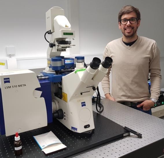
Dr Henri Franquelim is a Post Doc researcher at the Max Planck Institute of Biochemistry situated near Munich. He is a member of Professor Petra Schwille’s research team whose goal is to quantitatively understand the essence and core principles of living systems, with the very far goal of reconstituting self-regulating biomimetic systems in vitro. The overall research goals of the group are to investigate, under well-defined conditions, biological phenomena occurring at the membrane level, such as self-organization, pattern formation and membrane transformations. In order to do so, the laboratory utilizes various types of single molecule microscopy and spectroscopy techniques, employing in addition bottom-up reconstitution approaches with minimal components. Such strategies enable the study of selected subsystems under controlled and well-defined conditions, allowing the group to take full advantage of the strengths of single molecule techniques including atomic force microscopy (AFM).
Dr Franquelim’s work has applied AFM to study the remodelling aspects (controlling lateral segregation, bending, etc.) of biological lipid membranes. For example, he has been investigating the interaction of proteins, peptides and DNA origami nanostructures with lipid membranes; and to assess how such minimal components may self-assemble on top of membrane model systems. In a recent collaboration work with partners from the Chemistry Department at the Ludwig Maximilian University of Munich, Dr Franquelim further used the fast-scanning JPK NanoWizard® ULTRA Speed AFM to image with seconds/frame rates how photo-switchable ceramides upon photo-activation can reversibly modulate the structure of lipid raft-mimicking domains [1].
Asked about his motivation to use AFM for his research, Dr Franquelim said “Due to its sub-nm resolution and pN force sensitivity, AFM is a powerful tool to investigate model membranes. AFM grants us access to the topographical and mechanical properties of lipid domains and allows us to follow the interaction of biomolecules on top of such systems. Thus, the recent developments of fast-scanning imaging modes greatly boosted the biophysical repertoire of AFM. Now with the fast-scanning ULTRA Speed AFM from JPK, one is even able to follow at high-resolution the dynamics of proteins and other biomolecules on top of membranes, applying scanning rates near the ones used in conventional fluorescence confocal microscopy.”
The benefits for using JPK’s AFM systems date back to research at BIOTEC Dresden some ten years ago where Professor Schwille first combined AFM with fluorescence correlation spectroscopy (FCS) where JPK provided one of the first commercial AFM systems capable of operation on top of an optical microscope [2]. Dr Franquelim continued: “Nowadays (at the MPI Biochemistry), the group benefits in addition from the modularity of the JPK NanoWizard® systems, which not only offer a variety of very sensitive acquisition modes, but also permits the easy exchange of scanning modules between stations. The possibility to utilize the optical microscope while performing simultaneous robust high-resolution AFM measurements is the big advantage of JPK AFM’s. In this regard, we recently added the benefit of the JPK fast-scanning ULTRA Speed AFM system. This performs excellently at high speeds (0.5-10 s/frame), enabling us to follow and optically control dynamic events on top of model membranes. All-in-all, JPK’s instruments have proved to be very robust and user-friendly systems which are essential for the successful implementation of the group’s research goals.”
For more details about JPK’s AFM systems and their applications for the materials, life & nano sciences, please contact JPK on +49 30726243 500. Alternatively, please visit the web site: www.jpk.com or see more on Facebook: www.jpk.com/facebook and on You Tube: http://www.youtube.com/….
References
1. Frank JA, Franquelim HG, Schwille P, Trauner D (2016) Optical control of lipid rafts with photoswitchable ceramides. J Am Chem Soc, 138(39): 12981-12986.
2. Chiantia S, Ries J, Kahya N, Schwille P (2006) Combined AFM and two-focus SFCS study of raft-exhibiting model membranes. Chemphyschem, 7(11):2409-18.
JPK Instruments AG is a world-leading manufacturer of nanoanalytic instruments – particularly atomic force microscope (AFM) systems and optical tweezers – for a broad range of applications reaching from soft matter physics to nano-optics, from surface chemistry to cell and molecular biology. From its earliest days applying atomic force microscope (AFM) technology, JPK has recognized the opportunities provided by nanotechnology for transforming life sciences and soft matter research. This focus has driven JPK’s success in uniting the worlds of nanotechnology tools and life science applications by offering cutting-edge technology and unique applications expertise. Headquartered in Berlin and with direct operations in Dresden, Cambridge (UK), Singapore, Tokyo, Shanghai (China), Paris (France) and Carpinteria (USA), JPK maintains a global network of distributors and support centers and provides on the spot applications and service support to an ever-growing community of researchers.
JPK BioAFM | Bruker Nano GmbH
Am Studio 2D
12489 Berlin
Telefon: +49 (30) 670990 7500
Telefax: +49 (30) 670990 30
https://www.bruker.com/bioafm
Communications Manager
Telefon: +49 (30) 726243500
Fax: +49 (30) 726243999
E-Mail: bagordo@jpk.com
Talking Science Limited
Telefon: +44 (1799) 521881
E-Mail: jezz@talking-science.com
![]()
